

Computer-aided detection systems help radiologists spot suspicious patterns faster by marking areas in medical images that may deserve closer review. They do not replace trained readers. They act as a second set of eyes, especially in exams with subtle findings, high image volume, or repetitive screening tasks.
TLDR: Computer-aided detection, often called CAD, uses software to scan medical images for patterns linked to disease. In breast screening, for example, a CAD tool may flag clusters of tiny calcifications that could be missed during a busy reading session. In one screening center reading 200 mammograms a day, even a 5 percent gain in early suspicious finding detection can affect many patients over a year. The best results come when CAD supports, rather than overrides, clinical judgment.
Medical imaging creates a huge amount of visual data. A single CT scan can contain hundreds or even thousands of slices. MRI, mammography, ultrasound, and X-ray studies all add to the workload. Human skill remains central, but fatigue is real. Pattern recognition is hard work, and small abnormalities can hide in normal tissue. CAD software helps by drawing attention to image regions that match known warning signs.
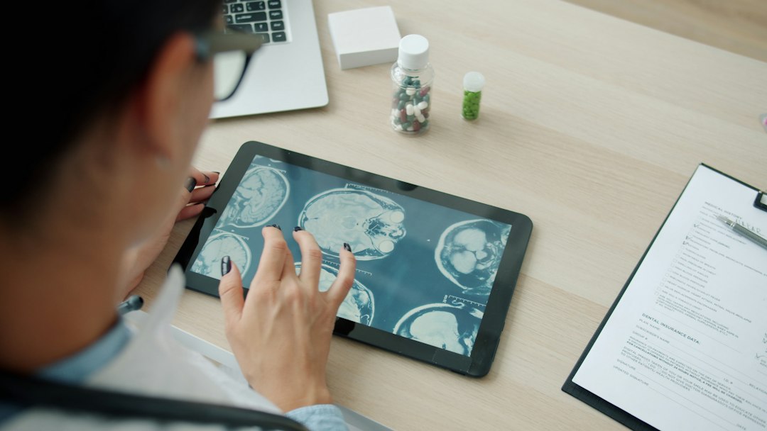
How CAD Systems Work
CAD systems analyze pixels, shapes, textures, density changes, and spatial relationships inside medical images. Older systems used hand-built rules. A lung nodule detector, for instance, might search for round objects with certain edges and density values. Newer systems often use machine learning and deep learning. These models are trained on large sets of labeled images, where known findings have already been marked by experts.
The software usually follows several steps:
- Image intake: The system receives an image from a scanner, PACS archive, or radiology workstation.
- Preprocessing: It adjusts contrast, removes noise, and standardizes image size or orientation.
- Feature detection: It searches for shapes, borders, textures, and intensity patterns.
- Risk scoring: It assigns likelihood scores to suspicious regions.
- Display: It marks possible findings with circles, boxes, heat maps, or overlays.
These marks are not final diagnoses. They are prompts. A radiologist still decides whether a mark is meaningful, harmless, or a false alarm.
Where CAD Is Used Most Often
CAD is common in screening and high-volume reading. It is used in several major imaging areas:
- Mammography: CAD can flag masses, architectural distortion, and microcalcifications.
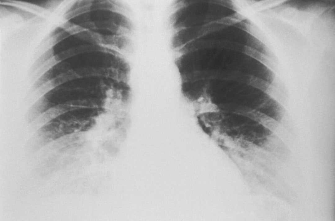
- Chest imaging: It can help identify lung nodules, pneumonia patterns, or signs of tuberculosis.
- CT colonography: Software can mark possible polyps inside the colon.
- Brain imaging: Some tools support stroke detection, hemorrhage alerts, and tumor follow-up.
- Bone imaging: CAD may help find fractures, density changes, or metastatic lesions.
In emergency care, speed matters. A tool that flags a possible brain bleed within minutes can help move a scan higher in the reading queue. In cancer screening, consistency matters more. The software keeps checking every case with the same level of attention, even at 4:45 p.m. on a packed schedule.
Why Pattern Detection Is So Useful
Medical images often contain tiny clues. A small lung nodule may be only a few millimeters wide. A faint breast calcification may blend into dense tissue. Early stroke signs can be subtle and easy to miss. CAD systems are built to notice repeated mathematical patterns that may be linked to disease.
That makes them useful in three main ways:
- They reduce oversight risk. The software can flag regions a human reader may want to inspect again.
- They improve consistency. The same rules or model are applied to every scan.
- They support triage. Urgent-looking cases can be moved ahead for faster review.
This does not mean CAD is always right. It means it can reduce blind spots. A tired radiologist still has expertise. The software adds a structured reminder system.
The Role of Artificial Intelligence
Modern CAD is closely tied to artificial intelligence. Deep learning systems can learn visual patterns from thousands or millions of examples. They may identify features that are hard to define with simple rules. For example, a neural network may learn the difference between a benign scar and a suspicious lung lesion by studying image texture, borders, and nearby anatomy.
Some AI-based systems also compare current images with prior exams. This is valuable. Growth over time can matter more than one isolated image. A nodule that was 4 mm last year and is 7 mm now needs attention. Software can measure and highlight that change. That saves time and cuts down on manual tracking errors.
Honestly, it feels like the most annoying part of some tools is not the detection itself. It is the extra clicks. If a radiologist needs 12 seconds to open a separate viewer for every flagged scan, that delay adds up across 80 or 100 cases. Good CAD should fit into the normal workflow, not slow it down.
Benefits for Clinicians and Patients
For clinicians, CAD can improve reading efficiency and confidence. It can help reduce variation between readers. It can also support junior staff by pointing out areas that deserve closer study. Senior radiologists may still reject many suggestions, but the marks can prompt a useful second look.
For patients, the main value is earlier detection and safer review. A suspicious finding found early may lead to faster treatment. A clean scan supported by careful human review and software analysis may also increase confidence. In screening programs, small improvements can scale quickly. If a hospital reviews 50,000 screening exams per year, even a modest improvement in detection or prioritization may affect hundreds of follow-up decisions.
Limits and Risks
CAD has limits. False positives are common in some systems. The software may flag scars, blood vessels, folds, dense tissue, or harmless shadows. Too many alerts can create alert fatigue. When every case has several marks, readers may start ignoring them. That defeats the point.
False negatives are another concern. If the software misses a lesion, a reader must not assume the scan is normal. CAD is support, not permission to relax. Training data also matters. A system trained mostly on one population, scanner type, or hospital setting may perform worse in another. This is a serious issue for fairness and safety.
Regulation and validation are also needed. Hospitals should check performance before full use. They should track missed findings, false alarms, report time, and patient outcomes. Claims in a brochure are not enough.
What Makes a Good CAD System
A strong CAD system should be accurate, fast, and simple to use. It should work inside existing radiology tools. It should show clear marks without covering key anatomy. It should provide useful confidence scores, not vague warnings.
Key qualities include:
- High sensitivity for clinically meaningful findings.
- Reasonable specificity to avoid a flood of false alerts.
- Seamless PACS integration with few extra clicks.
- Clear audit trails for quality review.
- Regular updates based on new data and clinical feedback.
The best systems feel almost invisible. They appear when useful, stay out of the way when not needed, and allow the radiologist to make the final call.
The Future of CAD in Medical Imaging
CAD will likely become more integrated with reporting, triage, and follow-up planning. Software may soon draft measurement tables, compare lesions across time, and suggest guideline-based next steps. It may also help predict risk, not just detect visible disease.
Still, trust must be earned. Hospitals need transparent testing, careful rollout, and ongoing review. CAD works best when clinicians understand both its strengths and its flaws. The goal is not machine control. The goal is better attention, faster recognition, and safer care.
FAQ
-
What is a computer-aided detection system?
It is software that analyzes medical images and marks areas that may show disease-related patterns. -
Does CAD replace radiologists?
No. It supports radiologists by highlighting possible findings. The final interpretation remains a clinical decision. -
Which scans can use CAD?
CAD can be used with mammograms, CT scans, MRI studies, ultrasound images, and X-rays, depending on the tool. -
Can CAD make mistakes?
Yes. It can miss disease or flag harmless structures. Human review is still required. -
Why is CAD helpful in screening?
Screening creates high image volume. CAD adds a consistent second check for subtle findings. -
What is the biggest weakness of CAD?
Too many false positives can waste time and distract readers. Poor workflow design can also slow reporting.